Retinal circulatory problems BRAO, CRAO, BRVO, and CRVO
The retina has three types of blood vessels: arteries, veins and capillaries. Blockage of a retinal artery is an inflow problem or occlusion or a stroke. Stroke does more severe and rapid damage. A stroke can happen in any part of the body, but we focusing on the retina. The blockage can occur in a small retinal artery and is called a branch retinal artery occlusion, and abbreviated BRAO. Blockage of the main retinal artery is called a central retinal artery occlusion and abbreviated CRAO and is the most severe. These are very serious problems requiring emergent attention. Blockage in a retinal vein is a problem with the outflow of blood and is not a stroke. More simply put, blood enters the eye without a problem but has difficulty getting out. Blockage of the main vein is called a central retinal vein occlusion, or CRVO. Blockage of a smaller vein is called a branch retinal vein occlusion or BRVO. These circulatory problems are serious, but not emergent like artery/inflow problems or occlusions.
The body and the retina have three types of blood vessels: arteries, veins and capillaries. Arteries have fresh blood loaded with oxygen and nutrients. Veins have used blood depleted of oxygen and nutrients. Capillaries are the smallest blood vessels and hook up the arteries and veins. Capillaries release oxygen and nutrients to the heart, brain, bones, muscles, skin, and every vital organs especially the retina. Artery blockage in any part of the body, is a stroke and always more severe. Retinal circulatory problems affect the arteries, veins, and capillaries in the back of the eye. Central retinal artery occlusion or CRAO and branch retinal art occlusions called BRAO represent blockage of the arteries or inflow of blood. They trigger more severe rapid painless vision loss and require emergent diagnosis and treatment. Central retinal vein occlusion, or CRVO and branch retinal vein occlusion or BRVO affect veins and capillaries. So retinal vein occlusion is an outflow problem and should not be labeled a stroke. CRVO and BRVO are also serious but associated with less severe vision loss and are not emergent. Unfortunately, patients cannot discern whether they have a retinal artery blockage or a retinal vein occlusion. Hence, time is critically important. Please act rapidly.
Symptoms to Watch For
The most common and critical symptom of all these retinal vascular occlusions is sudden, painless vision loss in one eye.
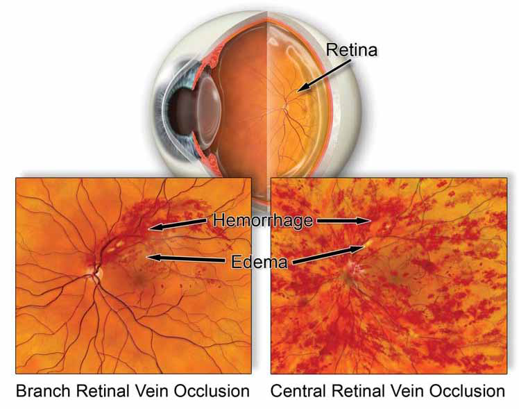
Severity Varies: The extent of vision loss depends on the size and location of the blocked vessel. Blockage of the main central vessels (CRAO or CRVO) typically causes more severe vision loss than blockage of a smaller branch vessel (BRAO or BRVO).
Other Symptoms: Patients may also notice a darkening, blurring, or a curtain-like shadow over their vision.
The Critical Distinction: Artery vs. Vein Occlusion
The symptoms alone often cannot tell a patient whether they have an urgent artery blockage (a stroke) or a serious vein blockage. This is why immediate medical attention is necessary.
How Is It Diagnosed?
The methods of diagnosis and treating CRAO, BRAO, CRVO, and BRVO must be individualized. Possible treatments could include specialized medicines injected in the eye, laser surgery, hyperbaric oxygen therapy, protocol treatments with blood clot dissolving drugs, and potentially a new experimental treatment with specialized red light therapy.
When to See a Retina Specialist
Please consult an eye MD and preferably a retinal specialist immediately if you have sudden painless vision loss in either eye. With retinal circulatory problems time is crucial!
Frequently Asked Questions
-
What causes retinal artery or vein occlusions?
While the immediate cause is a physical blockage, the blockages are often triggered by underlying systemic health issues. The most common risk factors for these occlusions include:
- High Blood Pressure (Hypertension)
- High Cholesterol (Hyperlipidemia)
- Diabetes
- Glaucoma
- Cardiovascular disease (especially for Artery Occlusions/Strokes)
- Smoking
- Certain blood clotting disorders
-
How will the doctor diagnose my condition (CRAO, BRAO, CRVO, or BRVO)?
Diagnosis is made through a comprehensive eye exam and specialized imaging. While the methods are individualized, they typically include:
Funduscopy: The specialist examines the back of your eye (the retina) to look for physical signs of hemorrhage or vessel swelling.
Fluorescein Angiography (FA): A special dye is injected into a vein in your arm, and photos are taken as it travels through the retinal vessels. This helps the doctor pinpoint the exact location and extent of the blockage and assess blood flow.
Optical Coherence Tomography (OCT): This is a non-invasive scan that provides highly detailed cross-section images of the retina to check for swelling and fluid buildup (macular edema)
-
Can vision loss from a retinal occlusion be recovered?
Recovery is highly dependent on the type of occlusion, the severity of the initial damage, and how quickly treatment is initiated.
Artery Occlusions (CRAO/BRAO): These have a poorer prognosis, as the retina is rapidly deprived of oxygen. Prompt, emergent treatment gives the best, though not guaranteed, chance of recovery.
Vein Occlusions (CRVO/BRVO): Vision may improve over time, especially with treatment to control swelling and prevent secondary complications like neovascularization (abnormal blood vessel growth). Treatment often focuses on stabilizing and improving the existing vision rather than full restoration.
-
If I have an occlusion in one eye, am I at risk of getting one in the other eye?
Yes. Since the risk factors for retinal occlusions (like high blood pressure and diabetes) affect your entire circulatory system, having a blockage in one eye signals an elevated risk for developing a similar problem in the other eye. Long-term management involves aggressively treating your underlying systemic health conditions to protect your vision in both eyes.

Don’t Wait — Protect Your Vision Now
Because a retinal artery occlusion (CRAO/BRAO) is a medical emergency requiring the fastest possible treatment, any sudden, painless vision loss must be treated as emergent until proven otherwise.
If you experience sudden, painless vision loss in either eye, please consult an eye MD and preferably a retinal specialist immediately.
Request an Appointment or call 208-535-4900 for immediate care at Teton Retinal Institute.